We assess a moving problem with a stationary snapshot, then wonder why two patients with identical arches respond completely differently to the same orthotic. This article lays out what static assessment does well, what it structurally cannot see, what only appears in motion — and when a static exam is still perfectly sufficient.
The short answer
- Static assessment captures shape and structure — arch morphology, alignment at rest, deformities, standing pressure. For accommodative devices and structural documentation, that's genuinely valuable.
- Static assessment cannot capture behavior — how the foot loads, rolls, and pushes off at real walking speed, or how it compensates when tired. Those mechanics are behind most of the symptoms patients present with.
- A dynamic foot assessment measures the foot doing its actual job. Modern tools capture 30+ biomechanical parameters during a ~3-minute walk in the patient's own shoe — no lab, no treadmill, no markers.
- The practical payoff: orthotics recommended from real gait data, and an objective before/after walk test that shows whether the device changed anything.
What static tools capture well — and honestly
Let's be fair to the foam box. Static and semi-static methods answer real questions:
- Morphology: arch height, foot dimensions, hallux valgus, claw toes, structural asymmetries
- Alignment at rest: rearfoot position in relaxed and neutral stance
- Standing pressure: a podoscope or pressure plate shows how weight distributes in bipedal stance
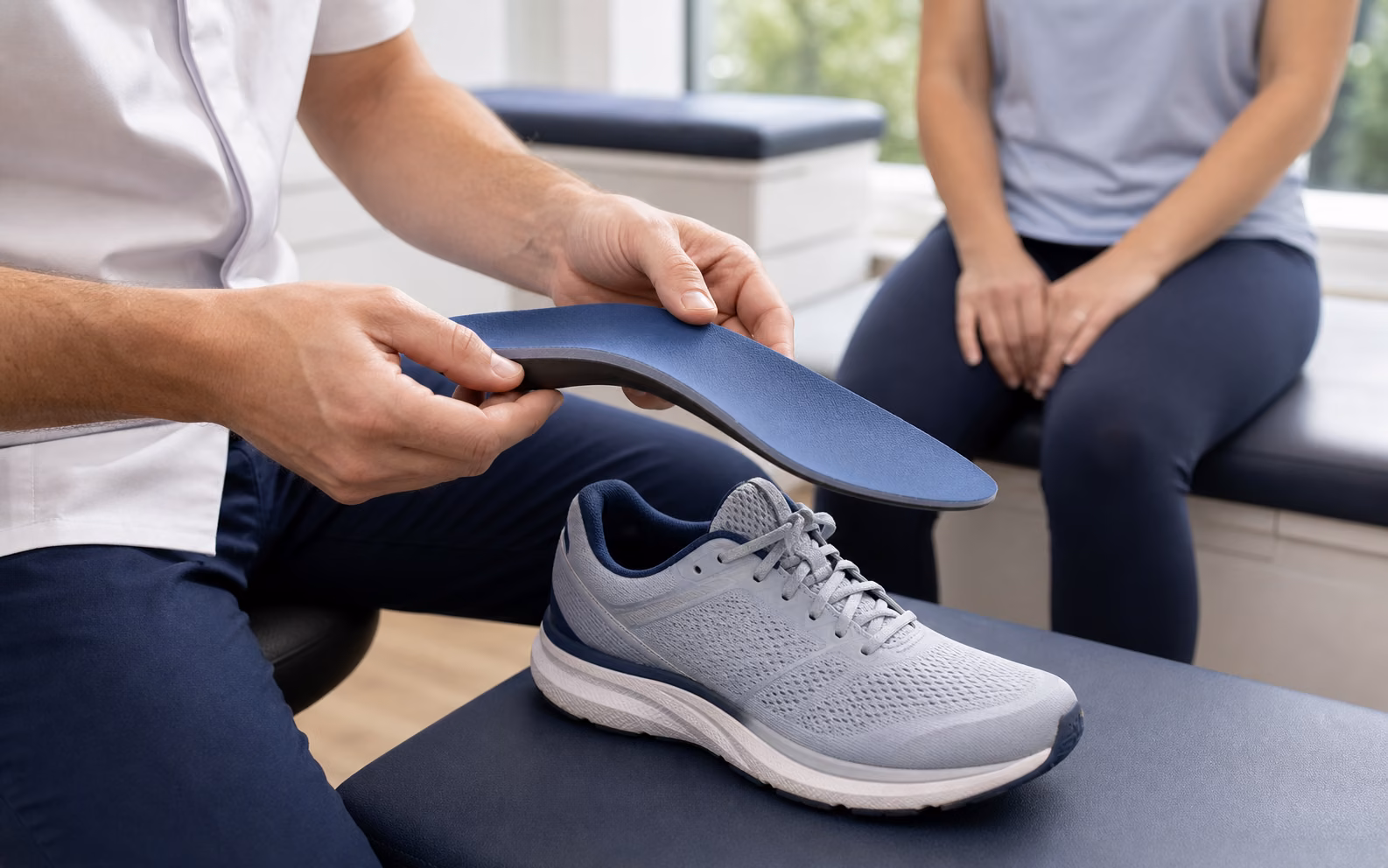
- A geometric mold: a foam impression or 3D scan gives the lab an accurate shape to build on
If the clinical goal is accommodation — offloading a fixed deformity, capturing shape for a diabetic patient's protective device, documenting structure — static capture does its job well. The trouble starts when we ask a shape measurement to answer a behavior question.
What the standing foot can't tell you
Standing still, the foot bears roughly half of body weight, symmetrically, with no momentum. Mid-gait, the same foot manages forces well above body weight, alone, in a few hundred milliseconds. Structurally, it's the same foot. Functionally, it's a different machine. A static exam cannot show:
- Loading in real time — where force actually concentrates at walking speed, which routinely differs from the standing pressure picture
- The roll-through — how the foot travels from heel strike to toe-off, and where that path deviates
- Push-off — propulsion strength and timing, and whether one side is quietly underperforming
- Compensation under fatigue — the pattern that only emerges after minutes of walking, which is exactly the pattern the patient lives in all day
- Side-to-side asymmetry in motion — two feet can look symmetric on a scanner and behave very differently in gait
None of this is a knock on the clinician. It's a limitation of the measurement condition: you cannot see loading behavior in a foot that isn't loading.
Findings that only show up in motion
Every experienced podiatrist has met these patients:
- The arch that collapses only under load. Normal-looking medial arch on the podoscope; at walking speed, the gait data shows it giving way mid-stance — precisely the mechanism straining the plantar fascia. The static exam said "normal foot."
- The asymmetric push-off. Recurring calf and Achilles complaints on one side. Shape: symmetric. In motion: propulsion measurably weaker on the affected side, with the other limb overworking to cover.
- The pain-free compensator. Knee pain, unremarkable feet. Dynamic data reveals an avoidance pattern in the roll-through — the foot is offloading an old, forgotten problem and sending the cost upstream.
- The fatigue reveal. A gait that looks clean for the first minute and deteriorates as minutes pass. A snapshot exam — even a short walkway pass — can miss what a sustained capture flags.
In each case, the finding that explains the symptom does not exist at rest. No foam box, however carefully pressed, contains it.
How a 3-minute dynamic capture works
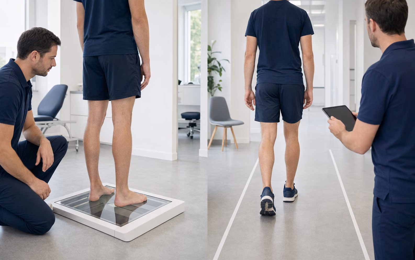
Dynamic assessment used to mean a motion-capture lab: markers, cameras, force plates, a research budget. That barrier is gone. In a Baliston-equipped clinic:
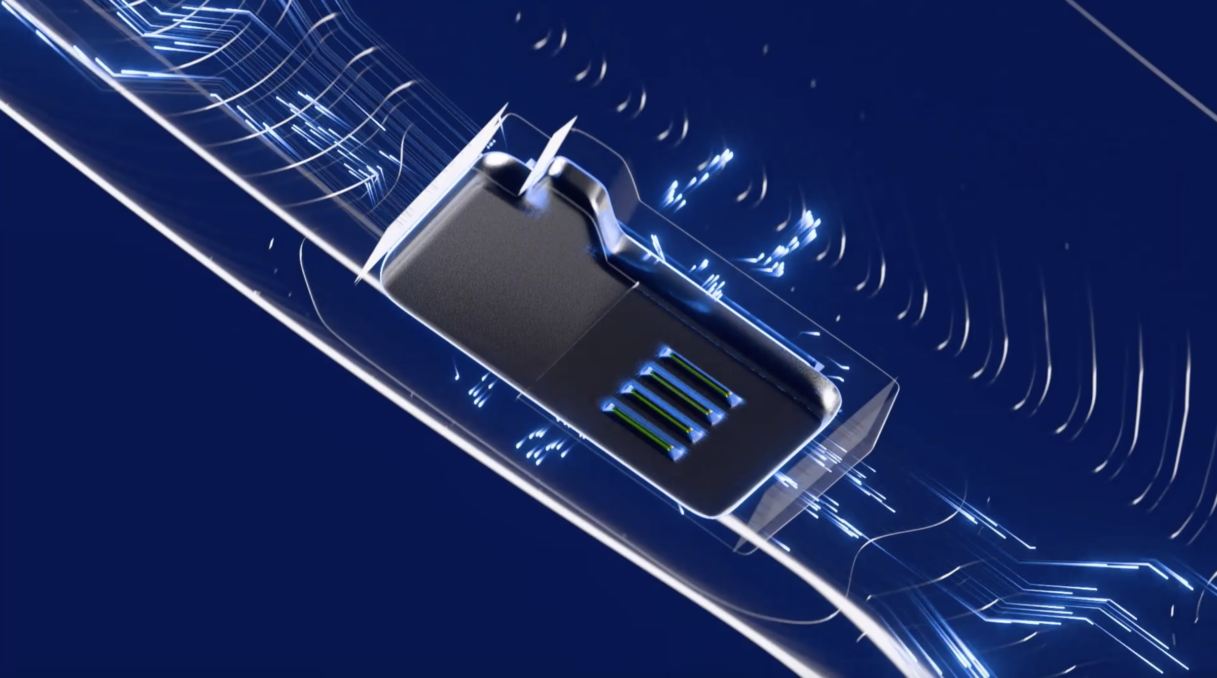
- The patient slips an insole equipped with AI Mov-Scan into their own shoe — the shoe they actually live in, which matters, because footwear changes gait
- They walk for about three minutes — a hallway will do, no treadmill required
- The system measures 30+ biomechanical parameters across the gait cycle: loading, stance and swing phases, roll-through, propulsion, symmetry — with 95% concordance with optical motion capture, peer-reviewed
- Results land in a Full Clinical Report, and Balia explains any parameter in plain language — to the practitioner or the patient. Balia explains and suggests; the practitioner decides.
Three minutes of walking replaces guesswork about what the standing foot would do in motion — because it simply measures it.
What this changes for orthotics
This is where the static/dynamic distinction stops being academic. In the Dynamic Custom Orthotics workflow:
- The recommendation is built from behavior, not just shape. The orthotic targets what the gait data surfaced — the collapsing arch under load, the deviated roll-through, the weak push-off — rather than inferring function from a mold.
- The device gets validated, objectively. The patient walks again with the orthotic: a before/after comparison in real data. Did loading redistribute? Did symmetry improve? You show it in numbers, instead of asking the patient to "wear them a month and tell me how it feels."
- Follow-up becomes measurement. At review, a new capture tracks whether improvements held — and flags when a device needs adjusting, long before the patient can articulate why something feels off.
For the clinician, this closes the loop that static-only workflows leave open: prescribe → verify → adjust, all against the same objective baseline.
When static is still enough
Dynamic assessment is not a religion, and honesty builds more trust than maximalism. A static exam remains a reasonable endpoint when:
- The goal is purely accommodative — offloading a fixed structural deformity where shape capture is the whole job
- You're documenting structure, not investigating symptoms — routine surveillance, footwear sizing, orthopedic records
- The presentation is acute and non-mechanical — an infection or a fracture doesn't need a gait workup
But when the complaint is pain, recurrence, or instability — anything that happens while moving — the assessment should include walking. The clinical logic is hard to argue with: measure the foot doing the thing that hurts.
The bottom line
Static and dynamic assessment aren't rivals; they answer different questions. Shape questions deserve static tools. Behavior questions — which is what most foot pain actually is — deserve measurement in motion. For decades that was a lab-only luxury; now it's a three-minute walk in the patient's own shoe.
The standing foot tells you what a patient is built like. The moving foot tells you what's going wrong — and, after the intervention, whether you fixed it.
Baliston-equipped podiatrists assess your feet in motion — about three minutes, in your own shoes — before recommending any orthotic, and verify the result with a before/after walk test.
Dynamic gait data in ~3 minutes, orthotic recommendations built from real movement, and objective before/after validation for every device — used by 1,500+ practitioners in 50+ countries, with Balia to explain any result in plain language.